Tadpole Laboratory

Discover how we started using tadpoles in neuroscience and learn about how we study them in our lab.


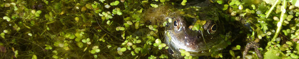
The Use of Amphibians in Research

The use of frog and amphibian tadpoles to study the brain began long before we started our lab.

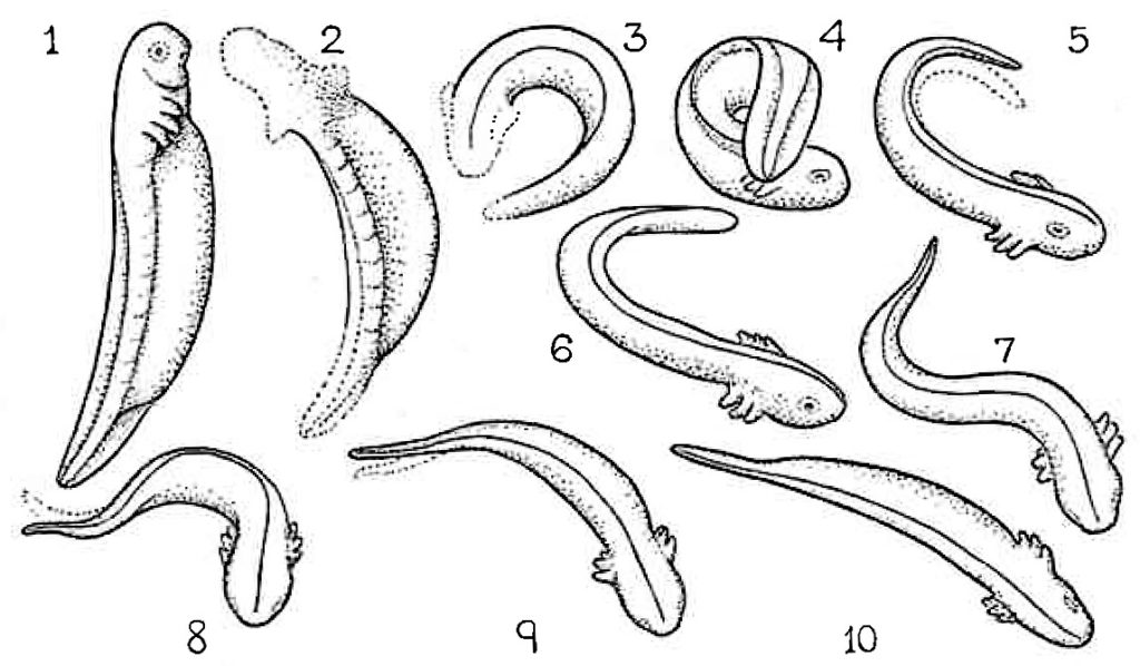
Coghill took motion pictures to define the development of the tadpole’s swimming behaviour in response to touch. It bends into a coil (1 to 4) and then swims off (5 to 10).

Xenopus laevis also are not the only species of frog that are used by neuroscientists and developmental biologists. While they are a popular animal model due to them being entirely aquatic and can be induced to mate using chorionic gonadotrophin, many other species of Xenopus can be used. To see the different species used in research, refer to Xenbase.


Producing Tadpoles

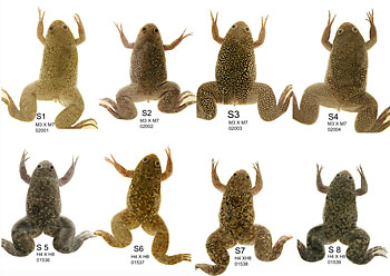
We have a breeding colony of South African Clawed Toads (latin name: Xenopus laevis). Each week we give a pair of adult toads an injection of the hormone human chorionic gonadotropin to induce mating and egg laying. Below is an image of what the adult toads look like, with females toads in the top row and male toads in the bottom row. We use their pigmentation patterns on their backs to recognise individual frogs.

S GROUPf
Adult Xenopus for breeding

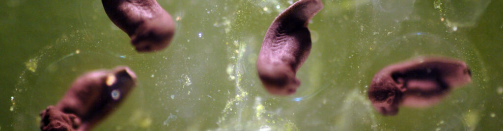
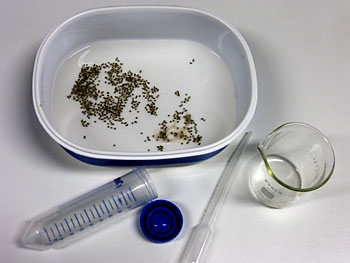
We collect the ~ 1mm eggs from the animal facility and after one day in the lab they look like this:

Eggs-in-dish-P1000990f
Eggs-in-dish-closer-P1000991f

The eggs are large (~1mm across) and as they divide to make an embryo with many cells, the yolk divides as well, so each cell has its own supply. This means that parts of the embryo can be removed and will survive and grow on their own. Small pieces can also be transplanted to new positions to see if they influence the development of other tissues (like the lens in the eye).

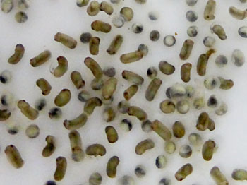
When you look more closely you can see that some are beginning to get longer and look like tadpoles. The round grey eggs are in the process of degrading.

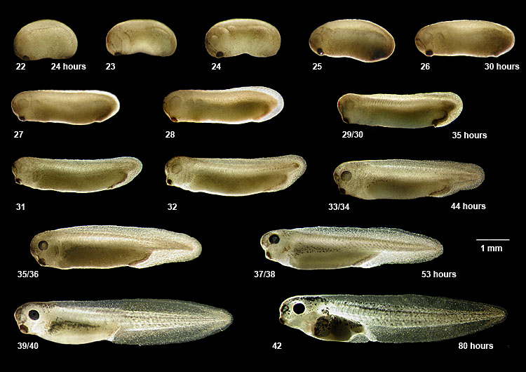
After 24 hours, the Xenopus embryo has made the beginnings of a nervous system. During the next 29 hours it grows into a tadpole and the nervous system and muscles start to function. At room temperature they reach the hatching stage after two days during stage 37/38 (see below). The image below shows the stages of development of a tadpole in the first three days after hatching.


The Tadpole Brain and Nervous System

tadpole-37-38-CNS-arrowsf

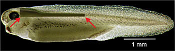
A newly hatched Xenopus tadpole is only 5mm long. Its brain contains about 3,000 nerve cells, called neurons. While the front section of the brain is only just starting to develop, most of the neurons are in the hindbrain and spinal cord, which is located between the red arrows in the image

The spinal cord is very small- about the same thickness as a human hair (~ 0.1 mm across)! The hindbrain (between the ears and above the gills) is bigger and has a fluid filled space or ventricle in the middle. The little yellow dots near the ventricle are neurons, which are ~ 0.01 mm across.

CNS-opened

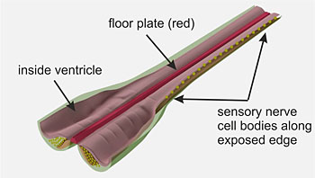
To expose neurons we open the hindbrain and spinal cord along the top, like shown in the diagram above. This immediately reveals sensory neuron cell bodies. These neurons have nerve fibres in the skin which are excited by touch, such as when the tadpole is prodded by a hair.

Slowed down video recording of a tadpole’s swimming response when prodded with a hair

By removing small areas of the wall of the ventricle, shown in pink the diagram, other types of neurons can be seen under a microscope. This allows us to record the activity of these neurons.


Recording Activity of Neurons

These small nerve cells communication with each other using electrical signals, which require specially made devices to record. When the spinal cord is opened, the tadpole is held on a small platform in a dish of salty solution (saline) to imitate frog’s blood. We put the dish on a powerful microscope with 400x magnification to see the cell bodies of neurons exposed at the opened top edge of the spinal cord. The long red arrow in the left image below shows where we position the dish:

recording-microscopef
Recording-electrode-on sensory neuron

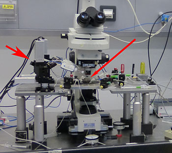
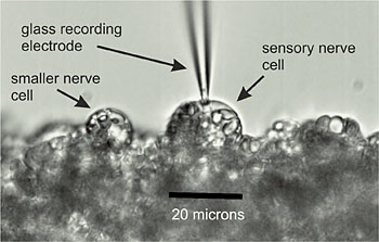
We use electronically controlled manipulators, shown by the short red arrow, to place a glass recording electrode onto a single neuron. In the picture above to the right, the electrode is placed on a sensory neuron. These are the largest neurons in the spinal cord and are excited when the skin is touched. A recording electrode allows us to record the voltage of the neurons we place them on, like the recording below:

Recording2

The neuron sits at its resting voltage and when the skin is touched, noted by the blue arrow in the image and time 0 in the graph, an electrical nerve impulse or action potential occurs. The voltage suddenly spikes for about 3 milliseconds.

This action potential is the electrical signal which travels along nerve fibres or axons so one nerve cell can excite or inhibit another at connections called synapses. That’s how electrical signals travel around the brain.


Recording the Connections Between Neurons

Nerve cells exchange signals at special junctions called synapses. To look for synaptic connection or synapses, we take recordings from two neurons at the same time. We stimulate neuron one by injecting a small current through its electrode to evoke an action potential and see how the other neuron responds.

In this case, there is a response in neuron two, which starts shortly after the action potential in neuron one. The delay is about 2 milliseconds.

synapsef

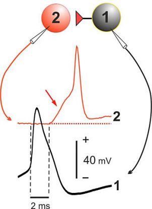
This small delay tells us that the electric current is not flowing directly from neuron to neuron. In reality, the following events occur to allow the electric current to travel from one neuron to the next:


The action potential in neuron one intiates the release of a chemical messenger, known as a neurotransmitter.


The neurotransmitter spreads across the synaptic gap to neuron two.


The neurotransmitter binds onto special receptor molecules on neuron two.

The activated receptor molecules cause channels within the neuron’s surface to open, so electrical current moves into neuron two and causes a positive increase in voltage called synaptic potential (shown by the red arrow in the diagram above).


If the synaptic potential is large enough, it will initiate an action potential in neuron two (shown in the diagram above).

This is how a signal is passed from one neuron to another in the nervous system.

To see the anatomy of the neurons after the recording, we use a chemical marker or dye and let it spread from the recording electrode into the neuron. We process it so the spread is visible under the microscope, which allows us to take detailed photos.

sensory and pathway neuronsf

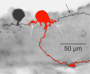
This photograph from one side of the spinal cord shows the sensory neuron one (shown in black) and neuron two (shown in red) from the recording we looked at above. White arrows show possible synaptic connections. The black arrow shows the axon or nerve fibre of neuron one.


The methods described here can be used to study many types of neuron in the tadpoles brain and spinal cord. For more details go to the Science pages.

The Digestive System


The digestive system is modified to account for the change of the herbivorous diet of the tadpole to the carnivorous diet of the frog.

This will close in 0 seconds

The Skin


The skin adapts for the change from a purely aquatic lifestyle to an amphibious lifestyle.

This will close in 0 seconds

The Reproductive System


The urogenital system develops to allow for reproduction in adulthood.

This will close in 0 seconds

The Skeleton


The skeletal structure develops to accommodate the change from tail swimming to using legs to move around. The skull also needs to be remodelled for a frog's change in vision.

This will close in 0 seconds

The Nervous System


A tadpole sees from eyes that are positioned on opposite sides of the head. During metamorphosis, the optical nerves develop to accommodate a frog's binocular vision, where the eyes are positioned at the front of the head.

This will close in 0 seconds

1. Mating and Laying Spawn


Male and female frogs go to ponds in the winter. They mate in the spring, and the female lays big clumps of eggs.

This will close in 0 seconds

2. Frog Spawn


Frogs eggs are called frogspawn. Each round black egg is about 1 mm wide and is surrounded by a blob of jelly. Other animals produce spawn as well, which you can look at here.

This will close in 0 seconds

3. Maturing Frog Spawn


After a few days, the eggs begin to grow into tiny tadpoles inside the jelly.

This will close in 0 seconds

4. Hatchlings


Then the tadpoles hatch! They are about 5 mm long and they can’t swim (yet). They can bend their body from side to side using special muscles along their trunks and tails.

This will close in 0 seconds

5. Young Tadpoles


When their tail is big enough, they swim off into the pond to start to feed. At first they have gills (the pale protrusions from the head region in the left photo) so they can breath underwater like fish. Young tadpoles feed by grazing the surface of pond weeds and also eating tiny floating plants called algae.

Click here to play a tadpole feeding game called Taddypole!

This will close in 0 seconds

6. Maturing Tadpoles


Later they develop lungs and can swim up to the surface of the water to breath. The gills are absorbed back into their bodies and eyes develop. Older tadpoles are then able to feed on small animals like young insects.

This will close in 0 seconds

7. Mature Tadpoles


The next stage in development is to grow back legs. Tadpoles during this stage need to eat meat in order to get the proper nutrients to grow.

If you are looking after tadpoles, be careful as they can eat each other if you don't give them meat to eat! Click here to learn more about how to look after tadpoles as pets.

This will close in 0 seconds

8. Froglet


Finally, tadpoles grow front legs and their tail shrinks until it almost disappears. This is when they climb out of the pond and start living on dry land. Small frogs are commonly called froglets.

This will close in 0 seconds

9. Adult Frog


The little froglets will stay by the pond and slowly grow over the summer, eating small insects and worms. They will hibernate just like other adult frogs in damp spots near ponds from autumn until the next spring.

After four years, the new frogs will become adults and will be ready to mate and begin the cycle again.

This will close in 0 seconds

Trial content.

This will close in 0 seconds

Copy - 9. Adult Frog


The little froglets will stay by the pond and slowly grow over the summer, eating small insects and worms. They will hibernate just like other adult frogs in damp spots near ponds from autumn until the next spring.

After four years, the new frogs will become adults and will be ready to mate and begin the cycle again.

This will close in 0 seconds

Desert Habitats


Desert habitats are the driest habitats in the world. Most people only think of very hot habitats as being deserts, but cold habitats can be deserts as well! Animals and plants that live in deserts have the ability to survive on very little water and animals can control their body temperatures so they stay at the right level.

 Some examples of plants and animals that live in deserts are cacti, the desert tortoise and the artic fox.

This will close in 0 seconds

Coral Reef Habitats


Coral reefs are found in warm tropical oceans all around the world. Coral reefs can be found in both shallow and deep water and take hundreds of thousands of years to grow! They provide food and shelter to many fish and other animals, making them habitats that are home to so many different types of life.

Some examples of plants and animals that live in coral reefs are the sea star, sea grass, the octopus and clown fish.

This will close in 0 seconds

Rainforest Habitats


Unlike deserts, rainforests are very damp habitats that are filled with lush plant and animal life. Rainforests have an average rainfall of 2,000 to 10,000 millimetres a year! That can be 10 times more rain that falls in the UK! Animals and plants that live in rainforests are used to the wet and humid environment and are able to compete with all the different animals and plants around them.

Some examples of plants and animals that live in rainforests are orchids, the poison dart frog and the hummingbird.


This will close in 0 seconds

Pollution of Habitats


Pollution is the contamination of habitats with harmful substances. These harmful substances can be anything from plastics, to fertilisers used in fields, to waste products from manufacturing factories.

As humans continue to reproduce and the global population grows, we continue to produce a larger and larger amount of waste and pollution. This affects all air, land and water-based habitats and threatens the health and survival of the plants and animals that live in them, including humans!

This will close in 0 seconds

Land Use


As the human population gets larger, we require more land to live on. More houses and schools and everything else we need to live must be built and more resources need to be found. As we expand our towns and cities, this takes away space and resources from other animals and plants.

In order to build more buildings for humans, land must be cleared, which greatly reduces its biodiversity. This means that the range of animals and plants in the land gets smaller, which can harm the food chains in the habitat.

This will close in 0 seconds

Deforestation


Deforestation is an example of harmful land use by humans. Trees are an important part of the carbon cycle and they help to clean the air and produce oxygen. With a growing human population, we have started clearing larger and larger areas of trees and forests to build on the land or to obtain wood.

This reduces the biodiversity the the habitats and sometimes destroys habitats completely.


This will close in 0 seconds