Rapid, innate and stereotyped animal reflex responses
- Nervous coordination of reflexes
- The involuntary reflex response
- The hatchling Xenopus laevis tadpole
- Rapid reflex responses in a hatchling tadpole
- The Xenopus laevis tadpole nervous system
- Summation
1. Nervous coordination of reflexes
In this section, we are going to consider how nervous systems allow animals to coordinate involuntary actions and react to stimuli.
Reaction to certain stimuli may mean the difference between life or death (or serious injury!) and therefore need to be fast.
For example, many animals have involuntary escape reflexes that allow them to escape quickly from predators or other types of dangerous stimuli.
These responses are normally rapid, innate reflex responses involving a short nerve pathway, allowing quick movement, usually away from the stimulus.
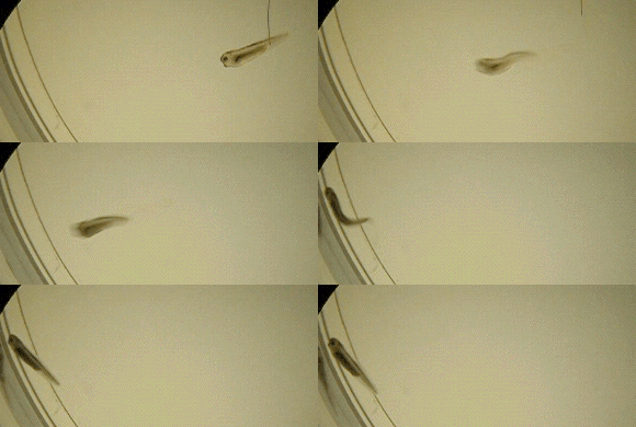
For example, when a tadpole is touched on its side, a reflex pathway is triggered and the animal flexes and swims away as seen in the video the right.
Before we consider how a reflex response is generated, first let’s recap on how a nervous system is organised:
A nervous system contains two regions:
The central nervous system (CNS), including the brain and spinal cord.
The peripheral nervous system (PNS), including the sensory and motor systems.
Sensory information is processed by the PNS and passed to the CNS where it is processed and sent back to the PNS in the form of motor information, for some kind of response.
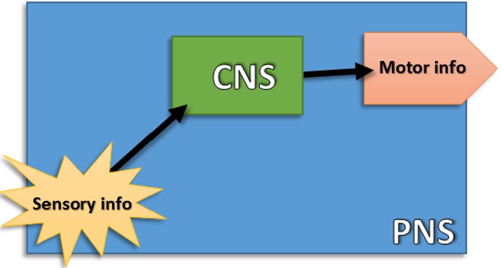
This motor response can be voluntary (involving decision making in the brain) or involuntary (involving no decision making and only the spinal cord).
As no conscious thought is required by the brain, involuntary responses (also called automatic or reflex responses) involve short nerve pathways and as a result, the time from stimulus to motor response is short.
These responses are often innate (do not require learning) and active from birth. They are therefore responses often key for survival.

A tiny hatchling tadpole needs to stay safe and swimming away from predators is crucial for its survival.
Therefore its developing nervous system has key pathways in place that allow rapid, innate behaviours. These include a touch reflex and a struggling reflex.
Click here for more info on the hatchling Xenopus tadpole. (hatchling story pop-up coming soon!)
2. The involuntary reflex response
Before we focus on the tiny tadpole case study, let’s first get to grips with the general background of reflex responses.
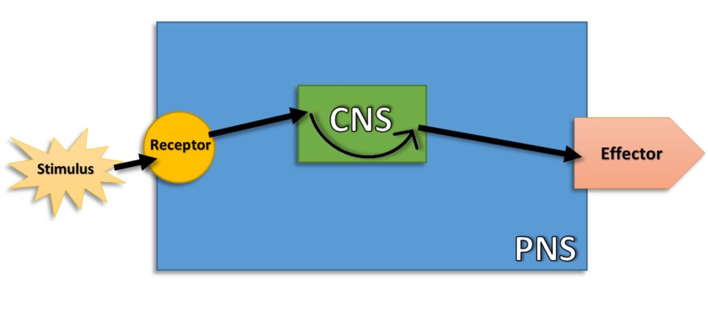
A reflex response is an involuntary reaction to a stimulus that results a response from an effector (usually a muscle or gland).
A stimulus is detected by a receptor, which triggers an action potential (AP) in a sensory neuron in the PNS.
This AP is carried to the spinal cord in the CNS by the sensory neuron along its axon, where it synapses with an interneuron(or relay neuron).
An action potential is generated in the interneuron, which in turn synapses with a motor neuron that has its cell body in the spinal cord of the CNS.
An action potential is carried from the CNS to the PNS along the axon of the motor neuron, which then triggers an effector to respond.
If the effector is a muscle, it may contract due to a release of neurotransmitter at the neuromuscular junction. If the effector is a gland, the gland may secrete something.
Examples often given in exam text books include the retraction of an arm when a hot stimulus is touched or the knee-jerk reflex.
However, with 86 billion nerve cells, the human brain is so complicated that even with decades of research by many dedicated scientists, we still do not understand the nerve circuits allowing us to carry out even the simplest of reflexes.
picture of complex nerve pathway coming soon!

3. The hatchling Xenopus laevis tadpole
One way to get around this difficulty in studying complex nerve pathways is to study simpler animals or indeed young animals that have not fully developed their nervous systems.
We can then begin to understand how specific neurons work together to produce a rapid, innate response, key to survival.
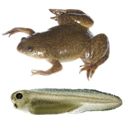
At the University of Bristol, we study the African clawed toad (Latin name: Xenopus laevis). In fact, we focus on tiny tadpoles that have just hatched out of their eggs. Only a few days old, these hatchling Xenopus laevis tadpoles provide a perfect animal model to study simple nervous systems.

Click here to read more about the hatchling Xenopus tadpole. POP-Up coming soon!
4. Rapid reflex responses in a hatchling tadpole
In this section we look more closely at a specific reflex pathway example. The concept is the same as you will find in text books, but with more specific examples. This section is meant to be an aid to understanding, not something to rote learn, so sit back and enjoy!

Hatchling tadpole
Xenopus laevis hatchling tadpoles of a few days old, have a simple nervous system and are able to react to stimuli they are likely to encounter in their watery environment.
Being small, numerous and nutritious, tadpoles are often the prey of a number of different animals and because of this, they need to be able to react fast to avoid predators.
Examples of tadpole predators:
Film of tadpole predators coming soon!
One rapid reflex response that the Xenopus hatchling has evolved is the flexion manoeuvre, so called because the body of the tadpole flexes when it bends away from the stimulation of the skin on its head or body.
The tadpole may then swim away in the opposite direction to the stimulus, which is an innate response to escape predators (see short film above).
In a reflex experiment in the lab, if the tadpole is touched very gently on one side of the body (at the little arrow), it bends away.
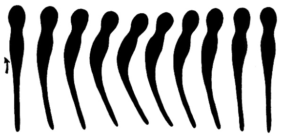
To the left is a series of stills from a high speed video (150 fps), showing a tadpole bending away from a stimulus on its body (represented by arrow).
The flexion of the hatchling represents a unique reflex example in neurobiology, as it is one where we know all of the neurons involved in the reflex arc and how the behaviour is generated.
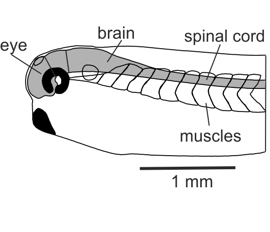
To the right is a diagram of the front end of a hatchling Xenopus laevis, including the nervous system, eye and muscles.
5. The Xenopus laevis tadpole nervous system
In this section we will look more closely at the hatchling Xenopus laevis nervous system and how behaviour, in response to a simple touch to the skin is generated.
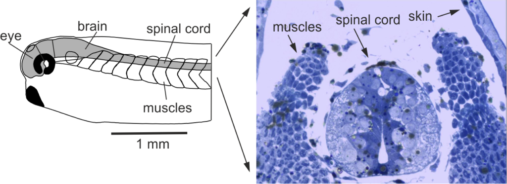
The Xenopus hatchling tadpole has a very simple brain and spinal cord (0.1 mm in diameter), with swimming muscles running down each side of the body.
If the tadpole is preserved and a slice is taken through the body and stained blue, we can see the skin, spinal cord and muscles on right and left sides.
Below is a diagram of the front end of a hatchling Xenopus laevis (left) and a cross section as seen under a microscope (right):
The sensory receptors that respond to touch in the tadpole are actually sensory neurons.
These sensory cells innervate the skin, which means their dendrites are close to the skin surface and are activated when the skin is touched.
To the right is a diagram of the spinal cord of the tadpole. Note the dendrite of the sensory neuron (yellow) is feeding information into the spinal cord in the CNS from the skin.
When the sensory cells are activated by the touch stimulus they fire an action potential, which is carried along their axons to interneurons (called sensory pathway neurons, in red),which in turn synapse with motor neurons (green).
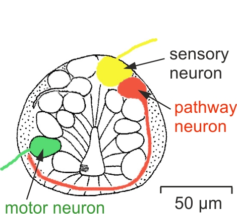
An action potential is then generated in the motor neurons, causing muscles on the opposite side to stimulation to contract. The tadpole will then pull its body away from the stimulus.

Left is the high speed outline of the tadpole flexion. Notice how the stimulus on the left side causes the muscles to contract on the right side, pulling the body away from the stimulus, before returning to the original straight position.
6. Summation
In this section we are looking even more closely into the flexion pathway in the hatchling Xenopus tadpole and the process of summation.
Summation is where input from a number of neurons causes a small amount of excitation (a small depolarisation) in another neuron and this excitation adds up to reach the threshold, triggering an action potential.
For a recap on action potentials and threshold voltages click here. (Pop-up box recapping action pots coming soon!)
Text books often imply that neurons synapse in a linear system, i.e. one sensory neuron, one interneuron and one motor neuron, and that the excitation (depolarisation) triggered in the post-synaptic neuron comes from one neuron only.
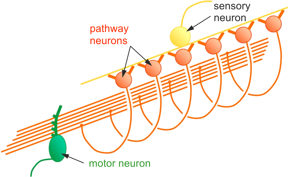
In fact, when one sensory neuron is stimulated, its axons may indeed run along the spinal cord and produce strong synaptic excitation of many interneurons.
This is the case in the Xenopus laevis hatchling flexion pathway.
In response to stimulation from a single sensory neuron, each of many sensory pathway neurons/interneurons fires an action potential and their axons cross to the other side of the spinal cord and make synaptic connections with motor neuronson that side.
However, each of these sensory pathway neurons/interneurons will only produce a weak excitation (a small amount of depolarisation) in the motor neuron and many will sum together to reach a threshold voltage.
If that threshold is reached, an action potential is generated in the motor neuron. This is called sensory summation (i.e. the summing together of excitation/depolarisation from a number of neurons all synapsing with one motor neuron).
The final part of the story is the response: When the motor neurons fire an action potential, the excitation or depolarisation travels down its axon and causes a neurotransmitter to be released at a neuromuscular junction. The muscles on the opposite side of the body to where the skin was stimulated will then contract, pulling the tadpole away from the stimulus.
Below is a diagram showing how a single sensory neuron (yellow) stimulates many pathway neurons (orange), which produce excitation in a single motor neuron (green).
For further information on how this process works click here. (link to further info coming soon!)

