Synapses

Structure and function of the junctions between neurons

  1. Synapses
  2. Synaptic tranmission

1. Synapses

Synapses are the junctions between neurons. They are structures where the terminal of an axon of one neuron meets (or synapses) with the dendrite of another neuron.

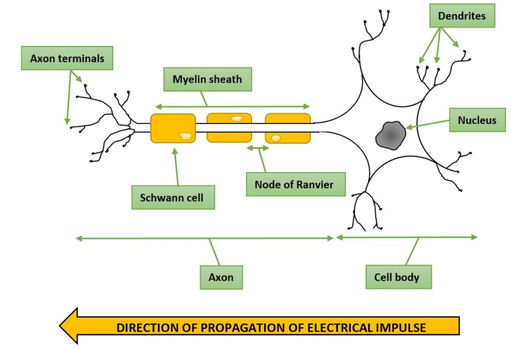
Below is a labelled diagram of a single neuron, indicating the way the impulse travels from the dendrites of the cell body to the axon terminals.
Next is a simplified diagram of many neurons synapsing with our original neuron.
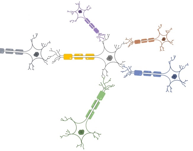
In reality not only can more than one neuron synapse with another, but more than one neuron can synapse to one dendrite.

There are two types of synapses- a chemical synapse (where a chemical neurotransmitter is released) and electrical synapsesor gap junctions (that allow current to flow through a pore from one neuron to the other).

But for now we are going to look at a typical chemical synapse between two neurons.

2. Synaptic transmission

When a nerve impulse arrives at the axon terminal of one neuron it is converted into a chemical message which is passed to the next neuron’s dendrite. Between them is a gap called the synaptic cleft over which the message is passed.

When we talk about the processes that go on at a synapse we refer to the membranes of these structures:

1. The membrane of the axon of neuron 1 is the pre-synaptic membrane (pre– meaning before, synaptic– of the synapse: i.e. “membrane before the synapse”) which is formed into the synaptic knob.

2. The membrane of the dendrite of neuron 2 is the post-synaptic membrane (post meaning after, synaptic– of the synapse: i.e. “membrane after the synapse”).

3. At a neuromuscular junction the post-synaptic membrane is that of a muscle cell called a sarcolemma.

Below is a labelled diagram of a synapse before an action potential/depolarisation has reached the synaptic knob.

The example is a cholinergic synapse, where the excitatory neurotransmitter acetylcholine is released.
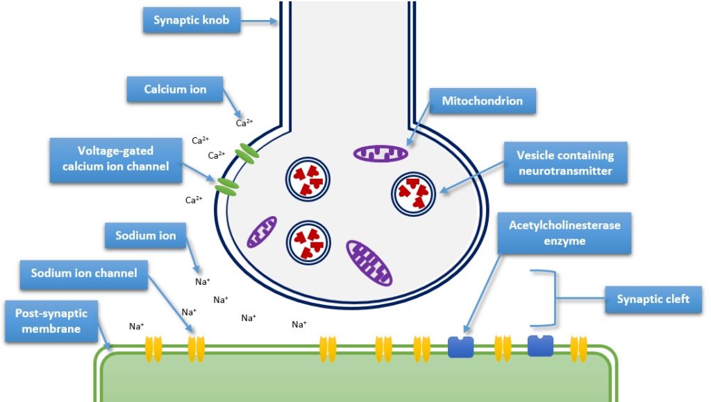
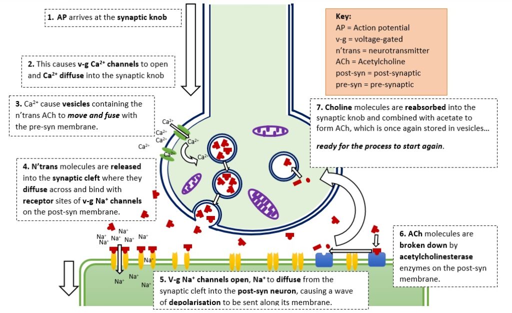
When an action potential (AP) reaches the axon terminal or synaptic knob a sequence of events is triggered that leads to a neurotransmitter (n’trans) being released into the synaptic cleft.

Below is a diagram detailing the key stages of the process at a cholinergic synapse.
The Digestive System


The digestive system is modified to account for the change of the herbivorous diet of the tadpole to the carnivorous diet of the frog.

This will close in 0 seconds

The Skin


The skin adapts for the change from a purely aquatic lifestyle to an amphibious lifestyle.

This will close in 0 seconds

The Reproductive System


The urogenital system develops to allow for reproduction in adulthood.

This will close in 0 seconds

The Skeleton


The skeletal structure develops to accommodate the change from tail swimming to using legs to move around. The skull also needs to be remodelled for a frog's change in vision.

This will close in 0 seconds

The Nervous System


A tadpole sees from eyes that are positioned on opposite sides of the head. During metamorphosis, the optical nerves develop to accommodate a frog's binocular vision, where the eyes are positioned at the front of the head.

This will close in 0 seconds

1. Mating and Laying Spawn


Male and female frogs go to ponds in the winter. They mate in the spring, and the female lays big clumps of eggs.

This will close in 0 seconds

2. Frog Spawn


Frogs eggs are called frogspawn. Each round black egg is about 1 mm wide and is surrounded by a blob of jelly. Other animals produce spawn as well, which you can look at here.

This will close in 0 seconds

3. Maturing Frog Spawn


After a few days, the eggs begin to grow into tiny tadpoles inside the jelly.

This will close in 0 seconds

4. Hatchlings


Then the tadpoles hatch! They are about 5 mm long and they can’t swim (yet). They can bend their body from side to side using special muscles along their trunks and tails.

This will close in 0 seconds

5. Young Tadpoles


When their tail is big enough, they swim off into the pond to start to feed. At first they have gills (the pale protrusions from the head region in the left photo) so they can breath underwater like fish. Young tadpoles feed by grazing the surface of pond weeds and also eating tiny floating plants called algae.

Click here to play a tadpole feeding game called Taddypole!

This will close in 0 seconds

6. Maturing Tadpoles


Later they develop lungs and can swim up to the surface of the water to breath. The gills are absorbed back into their bodies and eyes develop. Older tadpoles are then able to feed on small animals like young insects.

This will close in 0 seconds

7. Mature Tadpoles


The next stage in development is to grow back legs. Tadpoles during this stage need to eat meat in order to get the proper nutrients to grow.

If you are looking after tadpoles, be careful as they can eat each other if you don't give them meat to eat! Click here to learn more about how to look after tadpoles as pets.

This will close in 0 seconds

8. Froglet


Finally, tadpoles grow front legs and their tail shrinks until it almost disappears. This is when they climb out of the pond and start living on dry land. Small frogs are commonly called froglets.

This will close in 0 seconds

9. Adult Frog


The little froglets will stay by the pond and slowly grow over the summer, eating small insects and worms. They will hibernate just like other adult frogs in damp spots near ponds from autumn until the next spring.

After four years, the new frogs will become adults and will be ready to mate and begin the cycle again.

This will close in 0 seconds

Trial content.

This will close in 0 seconds

Copy - 9. Adult Frog


The little froglets will stay by the pond and slowly grow over the summer, eating small insects and worms. They will hibernate just like other adult frogs in damp spots near ponds from autumn until the next spring.

After four years, the new frogs will become adults and will be ready to mate and begin the cycle again.

This will close in 0 seconds